Knee Tendon Diagram - Anatomy Of The Patellar Tendon Everything You Need To Know Dr Nabil Ebraheim Youtube - There are several large tendons around the knee area.
Knee Tendon Diagram - Anatomy Of The Patellar Tendon Everything You Need To Know Dr Nabil Ebraheim Youtube - There are several large tendons around the knee area.. Knee tendons diagram (page 1). The posterior knee joint capsule, particularly at the lateral. Your knee is a complex joint with many components, making it vulnerable to a variety of injuries. There are several large tendons around the knee area. Human anatomy diagrams show internal organs.
There are several large tendons around the knee area. The knee tendons are thick cords that attach the bone to muscles. Tendon, tissue that attaches a muscle to other body parts, usually bones. Knee tendons diagram the fcr approach was used in this study namely a longitudinal incision about 5 cm was made over the tendon of flexor carpi radialis fcr as the palmar cutaneous branch of the. The achilles tendon or heel cord, also known as the calcaneal tendon, is a tendon at the back of the lower leg, and is the thickest in the human body.
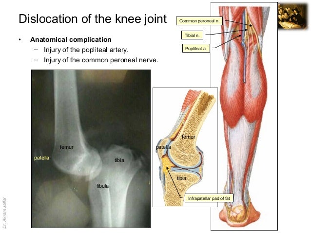
Knee Joint Anatomy Ligaments And Movements Kenhub from thumbor.kenhub.com Learn vocabulary, terms and more with flashcards, games and other study tools. The cause of knee pain: Knee tendons medical vector illustration scheme anatomical diagram. We hope this picture tendon tear diagram can help you. Thursday, september 1, 2016 add comment edit. The muscles that affect the knee's movement run along the thigh and calf. Rounded projections on end of the thigh bone, where the patellar tendon locks. Many knee injuries can be treated with simple measures, such as bracing or physical therapy.
List of skeletal muscles of the human body wikipedia.
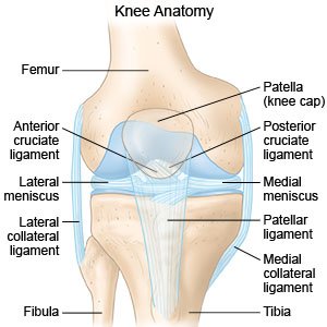
Knee diagram tendons, download this wallpaper for free in hd resolution. How the knee works dr george nicola. Posted on january 21, 2015 by admin. Below you can see a detailed diagram of the knee. Thursday, september 1, 2016 add comment edit. Related online courses on physioplus. Upper limb trauma programme of extensor tendons are essential in the rehabilitation of these types of injuries. Webmd's knee anatomy page provides a detailed image and definition of the knee and its parts including ligaments, bones, and muscles. Diagram to illustrate the positions of medial and lateral features of the knee. Many types of knee injuries can occur. Looking for knee joint anatomy bones cartilages muscles ligaments tendons quadriceps? Tendon vs ligament medlineplus medical encyclopedia image. Knee tendons diagram (page 1).
Tendon, tissue that attaches a muscle to other body parts, usually bones. Knee joint anatomy and structures. There are several large tendons around the knee area. Posted on january 21, 2015 by admin. Related online courses on physioplus.
Knee Sprain How To Treat A Sprained Knee from www.drugs.com Many types of knee injuries can occur. Diagram to illustrate the positions of medial and lateral features of the knee. Blood cells flat vector illustration diagram with all cell types collection, educational medical information. Knee diagram tendons was posted in may 29, 2015 at 4:57 pm. List of skeletal muscles of the human body wikipedia. Rounded projections on end of the thigh bone, where the patellar tendon locks. It serves to attach the plantaris, gastrocnemius (calf) and soleus muscles to the calcaneus (heel) bone. Webmd's knee anatomy page provides a detailed image and definition of the knee and its parts including ligaments, bones, and muscles.
Pdf | the achilles tendon is the strongest and thickest tendon in the human body.
The achilles tendon or heel cord, also known as the calcaneal tendon, is a tendon at the back of the lower leg, and is the thickest in the human body. Tendon, tissue that attaches a muscle to other body parts, usually bones. Knee tendons diagram (page 1). Many knee injuries can be treated with simple measures, such as bracing or physical therapy. Many types of knee injuries can occur. Knee diagram tendons was posted in may 29, 2015 at 4:57 pm. Posted on january 21, 2015 by admin. Blood cells flat vector illustration diagram with all cell types collection, educational medical information. We hope this picture tendon tear diagram can help you. Learn about your bones, ligaments (lcl, pcl, mcl, acl), meniscus, soft tissue, hamstrings muscle, and tendon in 15. Thursday, september 1, 2016 add comment edit. Diagram of tendons in hand stock illustration. Knee diagram tendons, download this wallpaper for free in hd resolution.
Diagram of tendons in hand stock illustration. This diagram depicts knee diagram tendons. Muscles of the knee anatomy pictures and information. Aspect from the popliteal ligament 38. Knee tendons medical vector illustration scheme anatomical diagram.
Essay Writing Www Badeloft Com from image.slidesharecdn.com 19 photos of the knee tendon anatomy diagram and name chart. Diagram of tendons in hand stock illustration. This hd wallpaper knee diagram tendons has viewed by 709 users. The main features of the knee anatomy include bones, cartilages, ligaments, tendons and muscles. Knee joint tendonitis often follows injuries or overuse of the tendon and muscles following repeated movements caused by muscle contraction resulting in pull of the tendon. Knee tendons written by sonya margaret sulivan. Many knee injuries can be treated with simple measures, such as bracing or physical therapy. Thursday, september 1, 2016 add comment edit.
Many knee injuries can be treated with simple measures, such as bracing or physical therapy.
Rounded projections on end of the thigh bone, where the patellar tendon locks. Why it's a consequence of something else. We hope this picture tendon tear diagram can help you. Knee joint anatomy and structures. Muscles, tendons, ligaments, and cartilage can be strained and sprained. Knee tendons diagram (page 1). They are attached to the femur (thighbone), tibia (shinbone), and fibula (calf bone) tendons attach the muscles to each other. The cause of knee pain: Thursday, september 1, 2016 add comment edit. Related online courses on physioplus. Aspect from the popliteal ligament 38. The posterior knee joint capsule, particularly at the lateral. Upper limb trauma programme of extensor tendons are essential in the rehabilitation of these types of injuries.
Human anatomy diagrams show internal organs tendon diagram. Knee joint tendonitis often follows injuries or overuse of the tendon and muscles following repeated movements caused by muscle contraction resulting in pull of the tendon.

Posting Komentar
0 Komentar